Ceramic rehabilitation of a worn dentition.
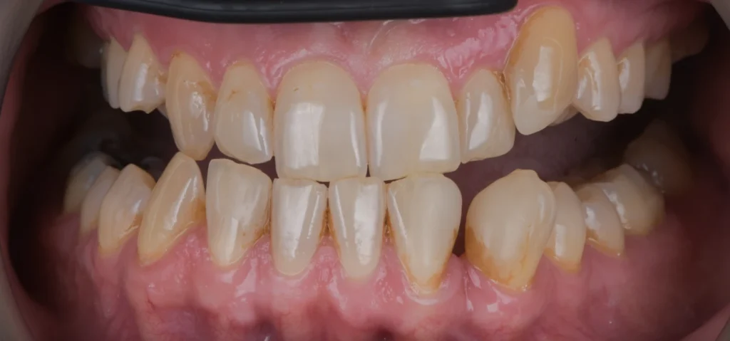
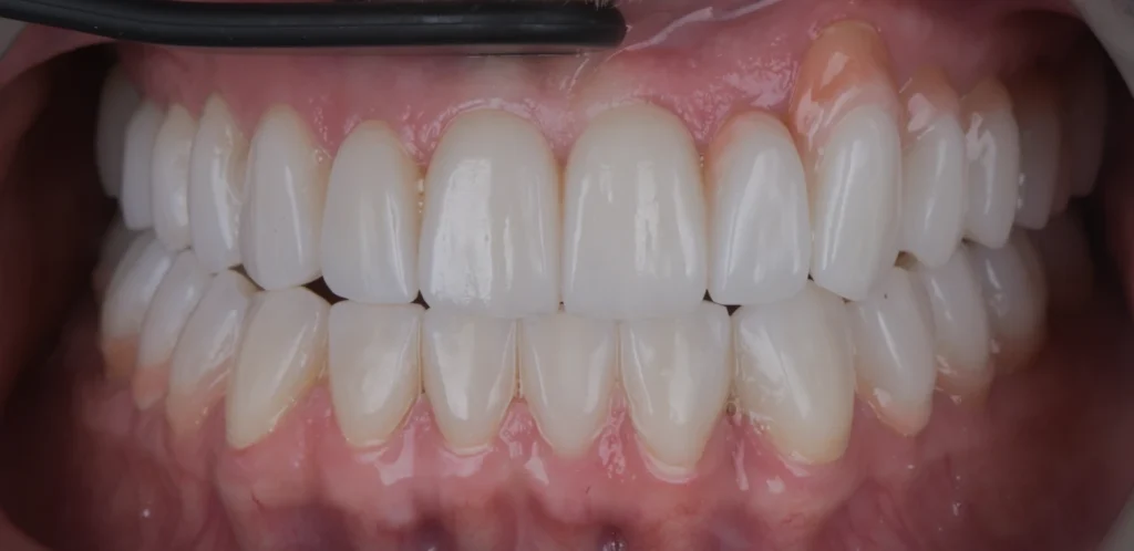
The pretreatment record shows irregular tooth form and uneven incisal edges. The post-treatment frontal photograph documents extensive ceramic restorative changes across both arches, with a more uniform anterior tooth form and a changed visible bite relationship.
What this illustrates +
Clinical assessment can examine tooth form and the visible relationship between the upper and lower teeth. Photographs alone cannot establish measured occlusal contacts, function, long-term stability or a legal conclusion.
Treatment by Dr Sahil Patel. Matching frontal views are shown at each clinical stage; the complete photographed frames are retained.